Essay
Six Weeks at Physio
A memoir from the 2026 MBL Cell Physiology course
From June 15 to July 26, I attended the Cell Physiology (Physio) summer school at the Marine Biological Laboratory (MBL) with 26 other PhD students and postdocs from all over the world. While the course had nothing to do with marine biology, we surely benefited from the ocean views and the diverse biological environment. We explored modern cell biology in depth using microscopic, biochemical, and computational approaches. The course was structured as 2 “skills weeks” followed by two 2-week rotations. In each rotation, three PIs would bring intentionally immature research projects from their labs, along with their graduate students/postdocs as TAs, to explore with us. Every morning there is also a seminar from 9, followed by discussions until noon. You might ask, what could one do in only 2 weeks? But we really did A LOT by sleeping a lot LESS! This memoir is dedicated to these 6 weeks. Partly to record the history, partly to preserve my perhaps immature and naïve scientific reflections on physics and biology.
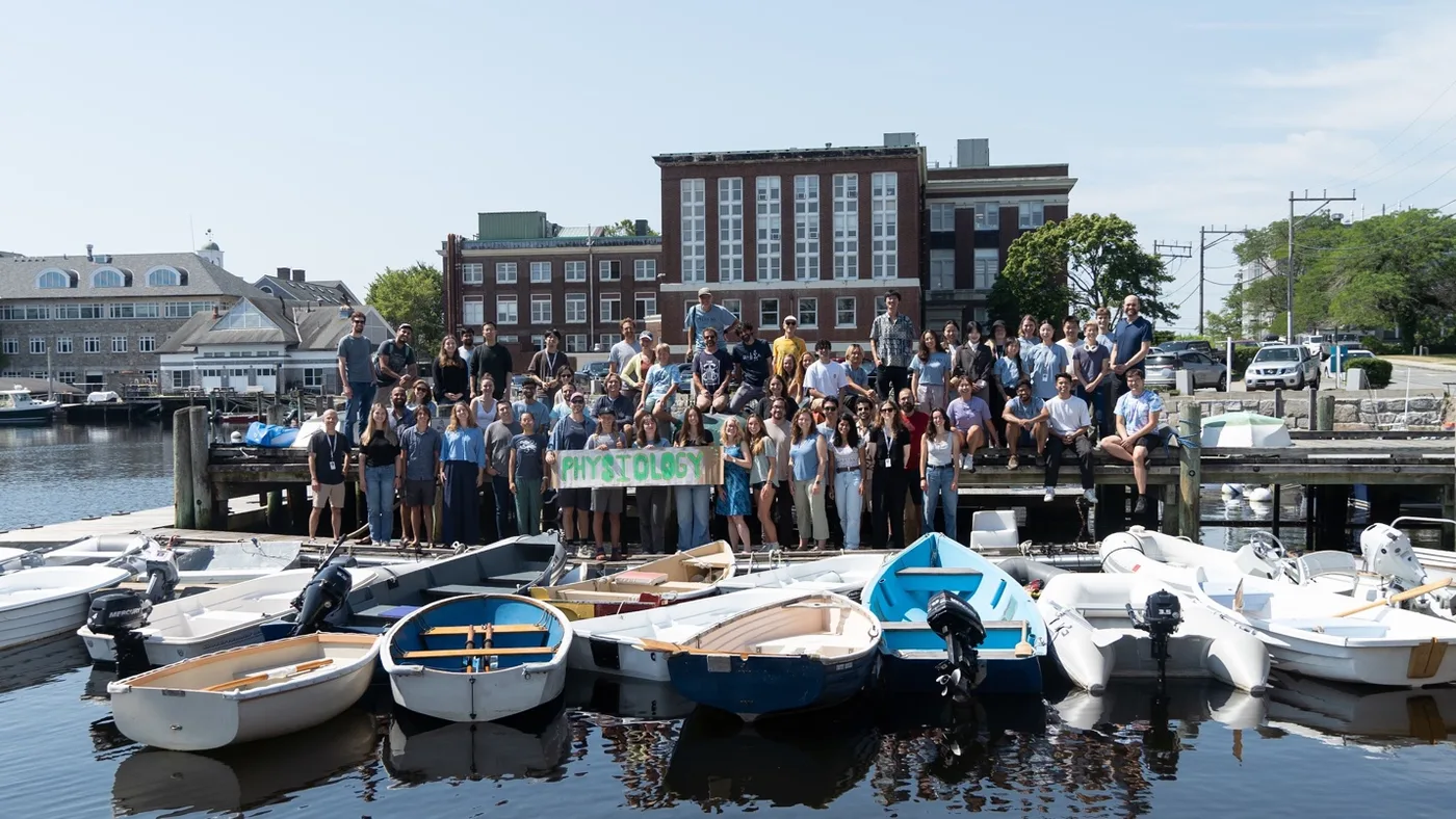
There are many fables of scientists who, after fleeing from reality to somewhere far away, released a magical gush of creativity and produced amazing work that had been totally unexpected. The example dearest to me when I was still an atomic physicist was Heisenberg’s flight to Heligoland, an island in the North Sea, in the summer of 1925 to avoid pollen. He returned to announce the birth of quantum mechanics. In cell biology, the 6-week Physio course has also been spawning such fables for the past century. MBL really has the perfect setting for this - next to the ocean, in a super-small town, and isolated from the rest of the world. When Jim Watson was teaching Physio in 1954, Meselson was his TA and Stahl was a student; at Physio, they began the “most beautiful experiment in biology.” In 1983, cyclin, the molecule controlling the cell cycle, was discovered during the course, work that later led to a Nobel Prize. Cliff Brangwynne, now the director of the course, first observed the formation of liquid condensates in cells while attending it in 2008. Since then, condensates have become a major theme in cell biology.
While no one could really anticipate such things happening, the overall expectations were high. There were multiple alumni of the course in my group, including my PI. According to them, the 6 weeks would be very intense, challenging, and forever life-changing. We would rest only on Sundays, when the major task would be to compensate for all the sleep deprivation from Monday to Saturday. I was certainly very excited long before I arrived. Having transitioned only the previous summer from experimental atomic physics to the theoretical study of 3D genome organization, I was still new to biology. My aim was to gain broad exposure to wet-lab techniques and develop intuition for what happens at the bench, so that I could collaborate better with experimentalists. But really, deep down in my heart, I wanted to become more like a biologist by attending this course and less of a physicist. Before joining my current group, a prominent biophysicist asked me to think carefully about whether I wanted to study beautiful physics or biology. Indeed, it is hard to imagine that the ultra-diverse biomolecular complexes in organisms would all follow some esoteric mathematical symmetry or field-theoretical structures. I wanted to study material, objective nature itself instead of carving out some far-reaching corners of Hilbert space, as people had been doing with exotic quantum matter in my previous field. I wanted to speak the biological language better by totally immersing myself in the world of cell physiology. Admittedly, there was a little bit of zeal of the convert.
Skills Week
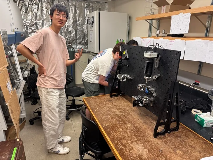
The first two weeks were the “skills weeks,” a series of 2–3-day modules combining lectures with hands-on work. In biochemistry, we dissected live squid and purified microtubules and kinesin. In microscopy, we built our own TIRF microscope from donated Thorlabs components, based on a design from Tanner, the man behind light-sheet microscopy. Then we looked at the samples we had purified with the microscope we had built! Very few groups actually saw microtubules gliding on kinesin with the home-built apparatus, although many could see it on a commercial TIRF. But still - good enough for a microscope built in 2 days! We stayed up until almost midnight every day, but there was also a lot of waiting between steps. Most of that waiting was filled with World Cup matches.
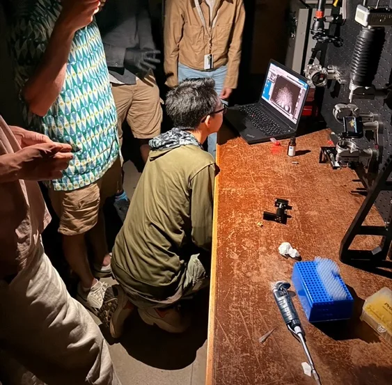
By then, David and Bob, who were fantastic biochemists and teachers, had just gotten me interested in learning more biochemistry. I wanted to know more about buffers, proteins, and all the other basic things. Then came the theory module. I was initially very much not excited. Most of the material came from Physical Biology of the Cell, which I already knew well from preparing for my oral exam. There was also 10% of me that doubted whether this could ever work, because calculus takes longer to get used to than pipetting. I would soon hate myself for thinking this way.
Christina and Deepak, the lecturers for this module, were Physio veterans who were now starting their own labs. Christina began as a biologist and gradually learned physics; Deepak went in the opposite direction. In only a few days, they covered basic statistical mechanics, probability, random processes, continuum mechanics, and my favorite, information theory! The depth came partly from forcing us to calculate on our own, which is still the most important part of learning any theory. But they also kept placing the mathematics back into biological contexts and relating it to phenomena that are basic enough but arouse immediate curiosity. They showed how mathematical language could convey our zeal and wonder about biological nature without becoming dry. Math usually scares people away. They made it intimate. The module climaxed with the estimation project, in which each group gave a 5-minute presentation in the style of a Fermi problem, also a format that kept recurring in our conversations afterward. I presented with Jan and Daniel on whether a flamingo diet would give us mercury poisoning before it turned our skin red!
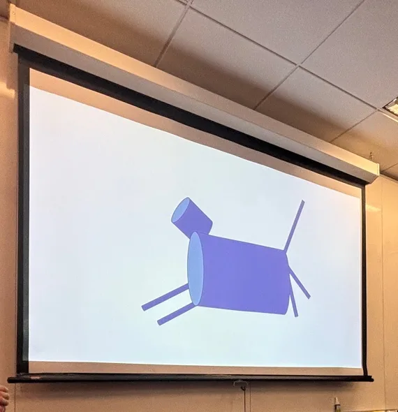
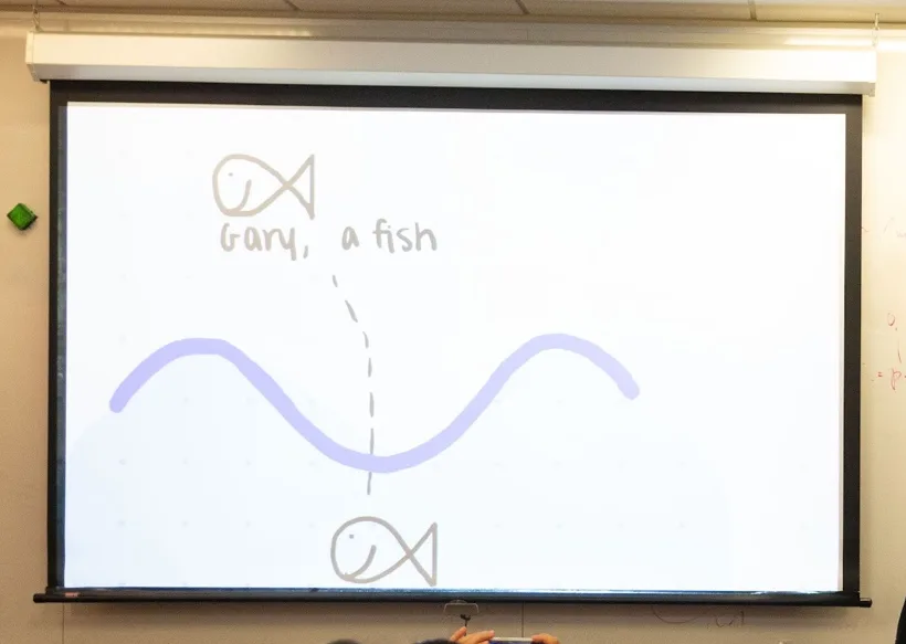
The influence of the theory module - and of Christina and Deepak - lasted throughout the course. Their account, in their final lecture, of how the course had influenced them was very touching. Their spirit was surely carried forward to the next generation of Physio: Mara and Karsh, my course mates who formed a similar pair - a physicist coming into biology and a biologist eagerly learning physics - later mimicked Christina and Deepak’s iconic laser-pointer toss in our rotation presentations. According to the rotation PIs, this year’s presentations contained much more modeling and theory than in previous years. Many course mates told me afterward that one of their biggest scientific inspirations from the course was to bring more theoretical thinking into their own work. After all, the biophysical principles underlying cell physiology are not quantum field theories. They are intuitive, intimate, tangible, and fun.
The omics module brought us back to the same squid: we sequenced RNA from the squid we had dissected earlier. At the end, Carry Albertin, a squid biologist, spoke about the still-understudied genetics of marine animals. She made me interested in the body plans of these non-model creatures.
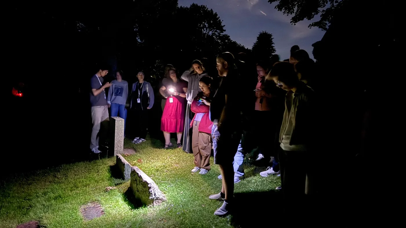
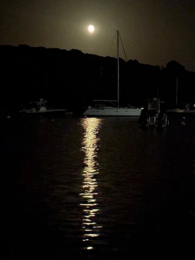
Following her talk, I immediately downloaded Hi-C data around the HOX genes of many different organisms. The gradual expression of repressed HOX genes during body-plan formation had been one of the original motivations behind my thesis project on 3D epigenetic memory. After staring at these maps for a while, I hypothesized that HOX genes had evolved from a more “1D-like” organization toward a more “3D-like” one as they became increasingly clustered in the genome across the tree of life, perhaps because a 3D epigenetic regulation can support higher plasticity if we treat development as a learning problem. I kept discussing this possibility with Carry and Yiyan during our class trip to the MBL graveyard that night. It was a fun trip; six Nobel laureates were buried there, bathed by the shiny moonlight, but I could not stop thinking and talking about my beloved HOX genes. Thanks to the course, the squid, and the Nobel laureates in the graveyard, I became a little more stimulated than usual.
The skills weeks ended with image analysis, computer vision, and AI. Beth patiently guided us through cell segmentation and the training of basic machine-learning models on our laptops. Gaudenz gave a series of talks that felt like farewell lectures. He was leaving academia for Roche Ltd., and the lectures became holistic reflections on everything he had engaged with over his career, from machine-learning algorithms to cell biology and causal inference. This is my favorite kind of talk: a rare chance to perceive someone’s scientific worldview as a whole. Pieces of knowledge can always be learned from ChatGPT, but it is the style of deep thinking that makes someone a unique scientist. I also love hearing hot takes, and Gaudenz was eager to delivered plenty. Out of respect for his strong distaste for social media and his request that the lecture material not be distributed, I shall stop here, except for one line that I am sure all of us will remember decades from now: “Do not ever, ever, ever, ever grow your cells on glass!” His eagle-like eyes might still secretly be watching over our cells…
Rotation 1
I was assigned to Cliff Brangwynne’s group for Rotation 1. Cliff had a personality very similar to my undergraduate PI’s, so I immediately felt somewhat close to him. In the spirit of Physio, I wanted to study something entirely different from my work at home: the nucleolus. In our group back at MIT, the nucleolus has always been regarded as very mysterious. We never talk about it. To us, it is mostly excluded volume inside the nucleus - a region the chromatin fiber cannot occupy. This is partly because we are a transcription-centered group, while the nucleolus prepares the machinery for the other half of the Central Dogma - translation. It does not translate itself; it transcribes ribosomal RNA and assembles the ribosomal subunits that will later carry out translation in the cytoplasm.
Unfortunately, I was initially not very motivated by my assigned project. It involved inducing cell–cell fusion to create artificial syncytia. Back at Princeton, Cliff’s group had been using osmotic shocks to change cytoplasmic protein concentration and study condensate dissolution, but the shocks stressed the cells. Cell fusion might provide a more physiologically permissive perturbation to tune the protein concentration in the cytoplasm. I was entirely uninterested in this avenue. To me, it was still fundamentally an in-vitro-style in-vivo experiment, i.e., still reductive phase-space-sweeping despite being in real cells. This still felt like condensate physics rather than fundamental biology, and again, I did not want to work on physics.
Another avenue they had conceived for this system was to use it as a tool to study the nucleolus. After 2 or more cells fuse, their cytoplasms mix, but their nuclei remain separate. The nuclei therefore live in the same cytoplasmic environment while retaining their original nucleoli - now forced, in a sense, to “communicate”. But I worried that the cell-fusion assay was quite destructive to the original cellular states, since it was inspired by viral infection. How physiologically relevant could the result be when the shared cytoplasm, cell size, and many signaling pathways might all change at once? I am not an immunologist; I wanted to study the nucleolus in “normal” cells. Still, I was very lucky to have Hailey, a graduate student in Cliff’s group, as my TA. She was kind, encouraging, and highly experienced, and saved me from many rounds of self-doubt in the coming two weeks.
We were asked to narrow down to a specific question by Friday, so that we could use the next week to explore it more deeply. My main goal was to narrow down what nucleolar “communication” really meant. Given the cell lines Hailey had prepared - there were really a lot! Ode to her detailed sample prep - we conceived of 2 possible directions. The first was material exchange. We would fuse a cell line overexpressing fluorescently tagged NPM1, the major scaffold protein of the nucleolus, with an unlabeled wild-type (WT) cell line. If NPM1 could leave the first nucleus, travel through the shared cytoplasm, enter the second nucleus, and join its nucleoli, fluorescence should gradually appear in the originally unlabeled nucleoli.
The second direction was to treat fusion as a rescue experiment. We would fuse a healthy cell with a cell in which RNA polymerase I - the enzyme that transcribes ribosomal RNA - had been acutely disrupted. The perturbed nucleoli could no longer transcribe rRNA normally and had an unhappy morphology. Would sharing cytoplasm with a healthy cell restore their transcription or morphology?
I felt very lost during the first week. The feeling that I understood too little about an artificial system struck me hard. For the entire week, my main goal was to get a handle on the most basic phenomenology of cell fusion. How does cell fusion happen spatially? How long does it take? What controls the size of the syncytium? (It was not until the end of the rotation that I realized that answering the “basic” questions I had listed earlier was itself a whole separate project, which was eventually what my very diligent lab partner, Anna T., pursued.) Most importantly, did the nucleoli show any signature of communication? If the answer was no, should I switch projects? In retrospect, I now understand that even if the answer had been no, it would still have been a valuable answer. Nothing is trivial or obvious in cell biology until you actually show it! So there was really nothing to worry about.
Physically, the week was also terrible. Woods Hole is next to the ocean, and the weather was quite unpredictable. Some days turned chilly under heavy thunderstorms. It was surreal to hold a pipette while rain driven by the howling sea wind pounded against the window. Due to the bad AC, those stormy days were also freezing inside the building.
The clock was ticking every day. There was not much room for me to read or think at the beginning, only an immediate dive into experiments. On day 2, Hailey, Nima (another TA), and I walked out of the tissue-culture room at 6 a.m. This was the cumulative outcome of my slowness in pipetting, my unmatched ambition with cramming too many fusion conditions into 96-well plates, and a temporary gas leak that got us kicked out of the building at 4 a.m. We returned 1.5 hours later. By the time we were done, the sun had already lit up Eels Pond, a beautiful water body right next to the lab. Then, after 2 hours of sleep, I felt 70% alive and prompted myself to keep going. I spent another entire day doing immunofluorescence for the first time, exhausted, only to realize that I had completed one-third of one page of the protocol. On the last night of week 1, I planned to collect some real data with a sample containing both protein tagging and RNA-labeling by 5-EU, which was supposed to give two separate readouts of structure and function. It became an absolute failure: for 3 hours, I kept finding and then losing focus on the goddamn spinning-disk confocal microscope. I felt so humiliated that I woke up at 6 a.m. to re-image it. Sadly, the results were not interpretable due to many reasons, some technical, some biological: over-expression causing subtle but non-trivial nucleolar state change, partially effective degron, uneven cell death rate confounding population level statistics, inability to trace back original cell line in fused cells, etc.
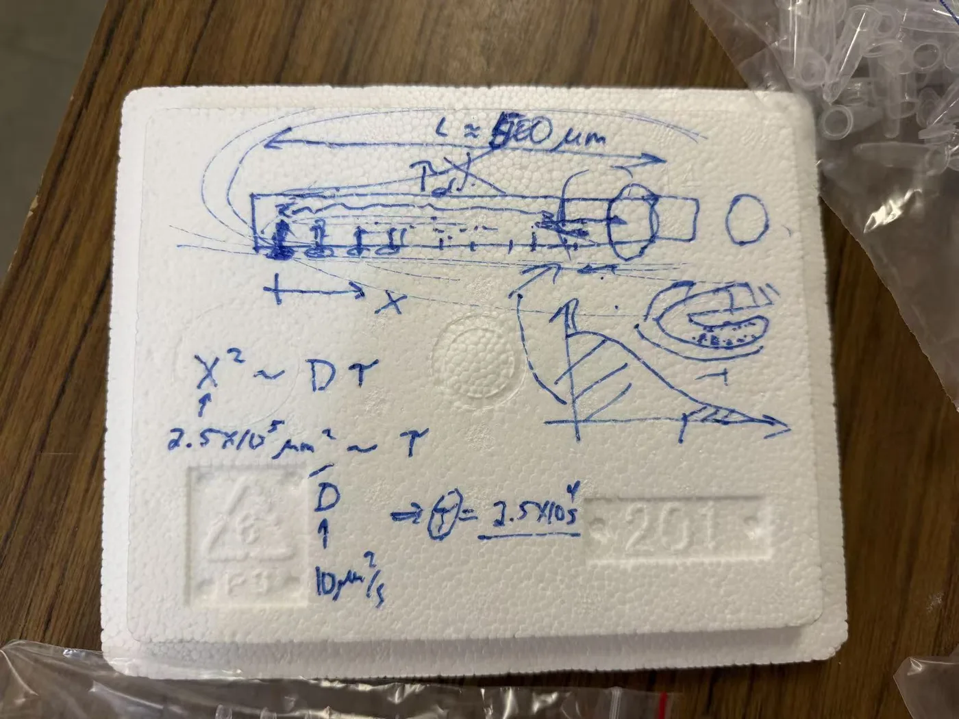
Amid all this confusion, one preliminary experiment did show something interesting. After I fused the fluorescent NPM1-overexpressing cells with WT cells, I did not see any fluorescence appear in the WT nucleoli, even hours after the cytoplasms had mixed. This suggested a total lack of movement of NPM1 and clearly violated the null expectation of concentration equilibration by diffusion. It should have rung alarm bells in us. Instead, I mostly treated it as a negative result for nucleolar communication - or simply as a sign that I had fucked something up.
While we could surely keep troubleshooting and do more experiments, they would be nothing more than extended random walking in the dark. Cliff also preferred to keep pursuing the NPM1 results, the big fish, rather than keep playing around with rescuing, because either outcome of the rescue would be equally unsurprising and equally uninformative. He had a point. It was finally time to think, and thinking helped a lot.
Ned Wingreen, a prominent theorist from Princeton, came to join us as our theory TA for the second week. While we were discussing my nucleolar-communication business, Ned said something about “ground state,” and a different question suddenly occurred to me: do intermediate nucleolar states exist? The nucleolus contains 2000 types of proteins, plus rRNA, so in principle its possible state space should be huge. Yet in cells we recognize only a small number of recurrent morphologies. Healthy nucleoli look broadly similar; when particular components are removed, the nucleolus often falls into another recognizable form, describable with phrases such as “GC/DFC inverted” or “cauliflower-like.” Why did such a complicated object occupy so few visible states? (I tried to compare this with the native state of protein folding. Both seemed to involve a high-dimensional search that nevertheless ended in a small number of reproducible structures. The analogy received some appreciation, although by pitching it I realized that the “native state” I had learned in my biophysics class was not quite the same object biologists had in mind. But what would be the chaperone for the nucleolus?)
Cell fusion now imposed some kind of coupling - a more general word than “communication” - between 2 nucleolar states through the shared cytoplasm. Would coupling generate new intermediate states, as in the case of an avoided crossing in matrix (quantum) mechanics? To sound even fancier: do nucleoli have identities? Are they plastic? I was satisfied that Ned, Cliff, and the TAs all seemed interested.
To test the idea, I fused WT cells with a TTF1-perturbed line whose nucleoli had a much more distinct morphology than the previous degron line. I then trained an autoencoder on nucleoli in the 2 unfused conditions. The purpose was straightforward: teach the model what the healthy and perturbed nucleoli looked like, then ask whether fused nucleoli resembled either parent or occupied new territory between them. Unfortunately, because of the limited time, I did not do a perfect job with cell segmentation and preprocessing. This made the autoencoder a little upset and prevented me from saying whether genuinely new states had appeared. At least the fused condition shifted toward more WT-like nucleoli, consistent with successful rescue. The larger question remained open, but I still thought it was solid and worth pursuing if my PhD had been about the nucleolus.
Meanwhile, we returned to the NPM1 result from week 1. I was advised to do a FRAP experiment on an individual nucleolus to show that the absence of transfer I observed earlier was not simply caused by NPM1 being sequestered within each nucleolus, forming aggregates. In that case, of course it could not equilibrate between nuclei. It would only be interesting when the lack of transfer persisted even though NPM1 exchanged rapidly between the nucleolus and nucleoplasm. Indeed, after FRAP, each nucleolus recovered within a minute, supporting my previous observations.
Even until the end, I still felt like doing essentially two very different projects at once and caught in between. The rescue project was ambitious but messy; the NPM1 project was narrow but much more tractable. To me, this is how research often feels: you have a difficult dream goal, but sometimes you also have an immediate low-hanging fruit that is equally important and must be addressed. I totally understood why Cliff and people were very excited about my second finding. It is the instinct of molecular biologists to search for new molecular mechanisms. The rescue idea was itself superficial to state. It was the underlying deeper object that was not well defined. What constitutes a nucleolar state, and what would count as retaining or changing identity? But that was precisely why it was exciting to me as a physicist. Physics (at least low energy physics), despite its heavy use of math, is fundamentally a subject of conceptual revolution, because experimental phenomena, math formalisms, and intuitive pictures are all unified as new concepts. We speak of (BCS) superconductivity as the “many-body interference of Cooper pairs”, not directly spelling out its wave-function. Probing intermediate nucleolar states is clearly along the lines of exploiting a new concept. I still felt the calling of a physicist.
Despite my internal struggle, the two projects sort of merged for the end-of-rotation 5-minute presentation. When cells were fused, their nucleoli could communicate, as evidenced by the rescue of function and morphology. Yet in our search for the physical substrate of such communication, we found that their material exchange might be regulated, at least for NPM1. As expected, the shape of the story did not follow our actual scientific progress at all.
Rotation 2
Christina and Deepak always said, “On the shoulders of giants,” referring to the authors of PBoC. I was assigned to work with one of these giants - Hernan Garcia. In principle, I should not have been assigned to him, since our group and his are equally transcriptional-centric. However, my project for this rotation was about transcription factors (TFs) which play key roles in transcriptional turn-on. In my research group, TFs have always been intentionally ignored and renormalized into an effective rate (k_on) in our modeling. In addition, all of our group’s collaborators grew cells on glass, whereas Hernan studies transcription in the context of embryo development. I was not at all a developmental biologist, but just as with the nucleolus, I expected to be transformed.
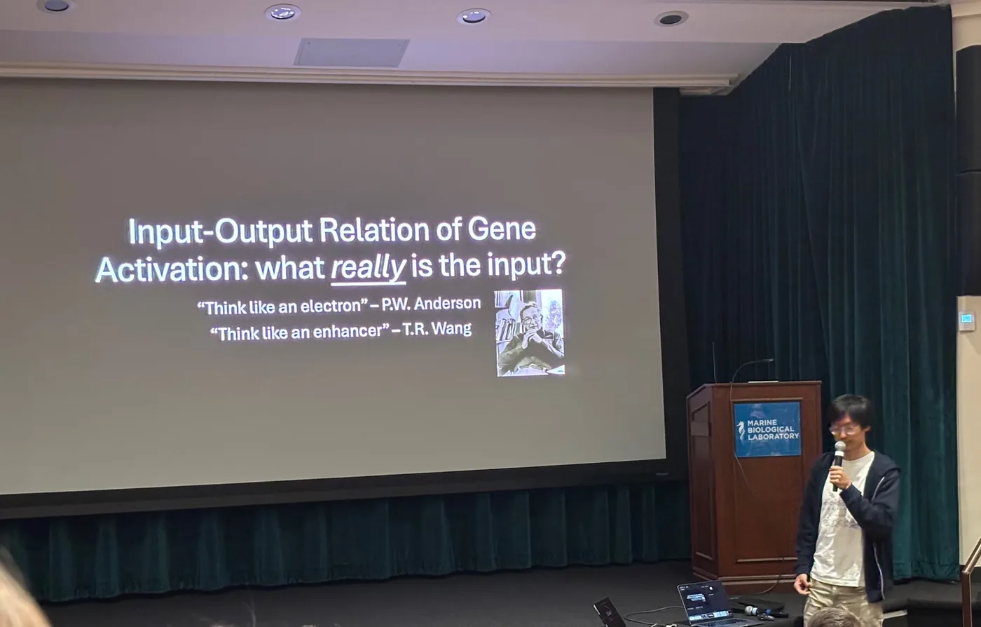
A classical question in the fly embryo is how smooth gradients of TFs such as Dorsal and Bicoid generate the development body plan with sharp boundaries. But another tentative project quickly caught my attention: the inhomogeneity of TF distribution inside a single nucleus. When we speak of the input-output relation between TF and transcription, what actually is the input seen by the gene? The usual formulation seems to assume one scalar value on the gradient curve. But what if the gene moves through a TF field that varies in space and time, so that its input is a highly fluctuating time series?
A short explanation of the system: The TF was Dorsal, but below I will simply call it “TF,” since dorsal also names a region of the embryo. Because the enhancer was immediately next to the promoter in our construct, I will address them interchangeably by the “gene locus.” We are using MS2 to look at nascent, fluorescent transcripts from the gene in real time.
I initially expected this rotation to be like a breeze (compared to the first one) because the questions are more well-defined, no cell work, and only two colors. Partly it was also because I had a fantastic TA. Aaron is a grad student in Hernan’s group and a person full of passion, curiosity, and warm encouragement. He really endorsed and supported me in exploring whatever interested me, unlike some other TAs who only made their students gather data for their own machine-learning stuff. Initially, I had the false impression that embryo experiments were easier than cell-culture experiments, since embryos were simply gifts from Mother Nature that could be taken for granted. In fact it was a significant underestimate of the challenges. Mounting embryos on coverslips was extremely difficult for me at the beginning. Though this time, those lying aphorisms came true. Failure did become the mother of success. I optimized the protocol a bit, found a previously unnoticed bug, and finally by the end of the rotation I became a mounting master.
The low photon counts were the biggest pain. It seems that nature likes to punish inquisitive scientists with ambiguous signals and a close-to-OK SNR that makes the hypothesis both right and wrong. This had also happened in my previous quantum-gas experiment. Part of the problem was imaging deep in z - it was an embryo, not a flat cell culture on glass! This posed stringent requirements on the microscope. I wasted some time on the wrong machine, but luckily learned a lot about microscopy thanks to Zach and Anna C., two microscope gurus from Vanderbilt. Eventually, the Nikon NSPARC was the only scope that could work, a super-res with enhanced detection for background correction. It was in the basement and had been flooded during Rotation 1, leading to several days of downtime. This time, I had to pray every day that it would not flood again and also fight with other groups when we signed up for microscope time for the next day. I was shamefully awarded the “police of NSPARC.”
The other factor for the low photon counts was the labeling. Adult flies ate Halo-dye-containing food and transmitted the dye to the embryo, where it labeled the Halo-tagged TF, so only a sparse fraction of TF molecules was visible. We had limited the dye because it was expensive. Then one day, we magically found a box in the freezer and started giving the flies twice as much. The photon counts did not increase. On top of that, there were only about 2,000 TF molecules in each nucleus. For comparison, one nucleus had roughly 5,000 voxels. Even before sparse labeling, there were fewer TF molecules than image voxels.
A detour on some theoretical conjectures: This 2,000-molecule problem is actually very interesting and fundamental to TF-mediated transcription. There are many more DNA-binding sites than TF molecules in a nucleus, so the sites must kinetically compete for a limited pool. This fact has historically given rise to the fundamental question of how TFs ever bind specifically to their own genes. How is this competition related to spatial inhomogeneity and chromatin-mediated cooperative binding, the two underlying contexts of my project? And most importantly, although a pure guess, could transcriptional bursting - a mystery that for decades - represent a reactive strategy that genes have developed in response to their “starving” situation? We know that the very non-trivial bursting statistics might not matter after all for the cell, because mRNAs are buffered at the nuclear membrane and when they travel outside, their statistics become Poissonian again. What kind of optimization problem does bursting correspond to? Aaron and I conceived of single-body pen-and-paper and many-body numerical simulations to address these questions, and I am very excited to run them after the course.
After fucking around for a while, we narrowed down our scope to a well-defined question rather than just asking “what is the input”: does TF concentration have to accumulate at the gene before transcription begins, suggestive of some kind of time-gating behavior? Intuitively, answering this question should be straightforward. Draw a small sphere around the gene-locus dot in each frame and count the total TF fluorescence inside it.
The experiments were straightforward. Data analysis was the true time sink. (I built a custom pipeline entirely based on the amazing previous work by a genius software engineer in Hernan’s lab, Yovan.) There is one small problem and one bigger problem. The small problem: no marker exists for the gene's position itself, only for active transcription. We got around this by exploiting the tail of a decaying burst - RNA Pol II molecules gliding off after a turn-off event left a residual mRNA signal that marked the gene's location just before its next turn-on.
The bigger problem is that, in most of the time, these nuclei were in G2 phase, so there were 2 copies of the same gene transcribing. Under the microscope, there would be 2 MS2 dots instead of one. Sometimes the 2 dots were clearly separated; sometimes they overlapped. Even within what the microscope called one frame, their relative positions could change across the sequential z-slices (we didn’t have a fast enough z-scanner installed unfortunately). It was therefore important to spatially resolve these 2 dots.
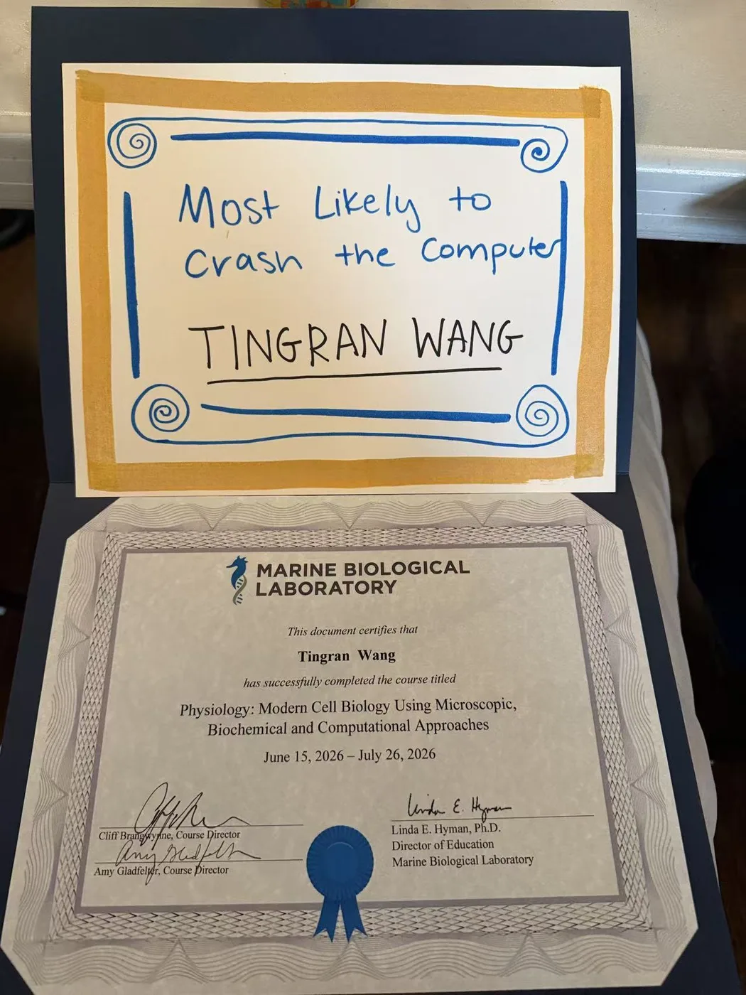
With Codex, I made an algorithm that reconstructed the 3D tube swept out by each sister chromatid dot during z-stack scanning. This would define a frame-rate-limited version of the local TF environment seen by an individual gene. In the data analysis parts of the day, I would often open a couple of Codex TUI sessions on the screen of the GPU workstation in the lab. Since not many people here use agentic AI, some people really wondered what the hell I was doing every day. Because my algorithm needed multi-threading and GPU computation, the fan would make some noise from time to time, which people quite vividly described as “airplanes taking off”. Together, my peers were very worried that I would break the computer. On the second to last day, I restarted the computer and all my previous work was wiped (the computer just crashed itself), causing me to pull an all-nighter to reconstruct my data analysis code through copied Codex chat histories. This led to on my Physio graduation certificate, I got awarded for the “person most likely to break a computer!” I guess that is what a true theorist could do.
The end results were mixed. On average, I found a very weak TF enrichment relative to the control. In some trajectories, TF concentration clearly rose before burst onset; in many others, there was no trend at all. I had taken far more data than the time allowed me to analyze, sadly. But data analysis in general was fun, especially with the guitar lullabies played by Garrison from the break room every night.
The power of observation
During one of our lectures, the lecturer asked who works on sequencing/omics experiments among our cohort. None raised their hands. “Cliff, you selected the right group of students!” he acclaimed, half-joke-half-seriously. This “dichotomy” between sequencing and imaging is of course totally artificial simply because there are amazing techniques like FISH and Optical Pooled Screening (which we were lucky to see the whole procedure, thanks to Nima!), but the core philosophy was still about direct observation under the microscope. It is the belief in the power of looking. No one can deny the immense power of sequencing and computation. But by the time a cell has been reduced to sequencing reads, genomic tracks, or an extracted time series, the cell itself - its morphology, motion, and all the things nobody thought to quantify, “the surprises” - has already made their exit.
“A good image is all you need.” This popular saying reflects the visual intimacy that only images possess. They are the natural attention seeker. Building this intimacy through prolonged looking under the microscope let us engage deeply with the cells in the most direct manner. In my rotation 2, I needed to first demonstrate before anything else that the TF concentration in embryo nuclei was indeed inhomogeneous. However, due to the low photon count elaborate above, in the raw data everything looked like pure noise. I decided to use a spherical-harmonic decomposition to show the inhomogeneity. To physicists, this would be quite natural; my biologist TAs and cohorts did not like it, mainly because they are not convinced by my control group, i.e. how I ran the same algorithm on the same data but randomly shuffled. Soon I learnt a much simpler route than this plumbing. There was a fantastic “trick” - maximum z-projection - that immediately made the inhomogeneity extremely visible. I was playing with this on Fiji, and Aaron, sitting 5 meters away from my screen, shouted “that looks non-uniform!”. Moreover, these features after max-z last over several frames, indicating they were true signals instead of statistical flukes. One might still object that this is just some trick that sounds like cheating, but the point is that I was not trying to systematically characterize the concentration field. I only needed to have a rough sense of the inhomogeneity features’ timescales and length scales, for which this was the most perfect. A good image gives you the “sense” of what you should even be hunting for.
Besides merely massaging the embryo images to test our hypothesis at the computer, we also all watched the whole process of embryo development while acquiring the data. It was mesmerizing. For example, we saw that nuclei falling behind in the synchronized wave of mitosis would quickly dissolve – like some error-correction mechanism - leaving a permanent “defect” in the original periodic arrangement of nuclei! These whole embryo level phenomena might not immediately relate to transcription, but once you move away from cell culture, every problem is fundamentally developmental. I would never have appreciated this if I had only looked at single nucleus, or time series downloaded from other papers. On the same note, my lab buddy back at MIT was also looking at MS2 signals to study transcription dynamics, though in mouse cell cultures. He was doing highly complicated statistical inference using time series already fully processed by his experimental collaborator. But now he decided to look at the raw movies, and it opened new worlds for him. After staring for long enough, we saw many interesting characteristics of transcription dynamics outside of our mental model. We started to know the system in a way that was difficult to acquire from the processed traces alone.
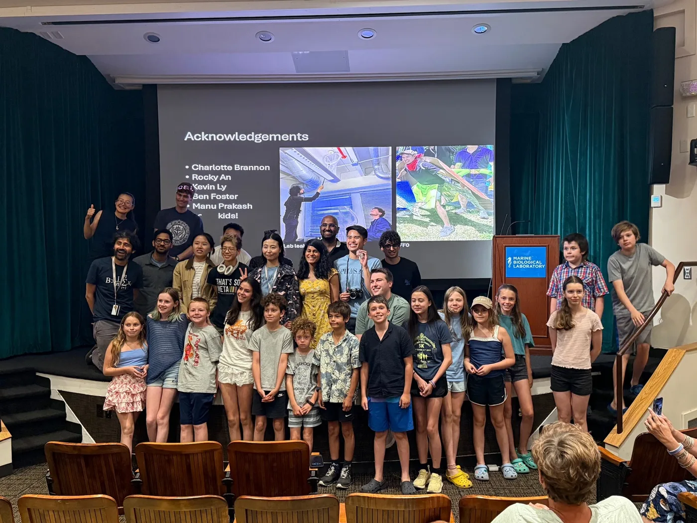
For Manu Prakash, a professor at Stanford who led a group in rotation 1, building this intimacy through microscopy characterized a major part of his entire scientific life. He is a Miyazaki-type of scientist: He respects nature, goes on fierce voyages to capture the weirdest creatures in the oceans and then systematically study them. I really appreciate how he loves these “recreational biology” (in his own terms) just for the sake of them, not because they can help cure cancer or they contain or break some exotic symmetry (many-body physicists know what I am talking about). Both in his morning seminar talk, and his students’ end of rotations presentations, there were many breath-taking pictures of non-model organisms that keep stirring human imagination. During rotation 1, I remember him sitting in the middle of the room, moving from one strange organism to another and explaining all the facts about each of them forever. We were all deeply impressed. One of his rotation projects was about capturing fireflies. They posted fliers on the street for 2$ per firefly captured. At the end-of-rotation presentation, they invited all the kids who participated. It was very touching.
Doing microscopy is about acquiring information. A microscope itself is essentially an information channel, transforming biological samples that store physical information (“information is physical!”) into digitized NumPy arrays. On a side note, in this view, Gaudenz also provided the best angle I’ve known to understand fixed-cell super-resolution microscopy like STORM, because it transfers the unused information in the time dimension into spatial dimension. Before microscopy has turned quantitative, it was mostly used to support verbal arguments or descriptions about cells. For that, people would make beautiful drawings from what they see through the lenses. Now, with technology advancements, we started to appreciate more and more the richness of images. An image often contains far more information about cellular state than we initially may know how to extract.
In a morning seminar, our speaker, Manuel Leonetti, talked about how label-free imaging can outperform single-cell RNAseq in predicting the phenotypes of cells. A surprising win – no staining needed since its label free, and also considering the dramatic cost difference One of our rotation PI, Lucas Pelkmans, developed high-throughput iterative Immunofluorescence assays for subcellular protein multiplexing, and achieve prediction of cellular states and observables only limited by physical limit (Poisson/shot noise, or measurement noise.) He transformed what people typically thought as “noise” or “cell-to-cell variability” into highly useful signals. Indeed, the differentiation between signal and noise is itself largely arbitrary, up to one’s scientific question. Until reaching true physical limit, latent variables should still be regarded as true information, not “noise” which sounds like black-box oracle word in this context. As technology progresses, we would surely be able to extract more and more information from images.
From staring at one cell, one embryo or one stentor for hours, to profiling millions of fixed cells, the scale was completely different. But the faith was the same: keep looking, because the image probably contains more than we currently know how to ask from it.
Fun outside science
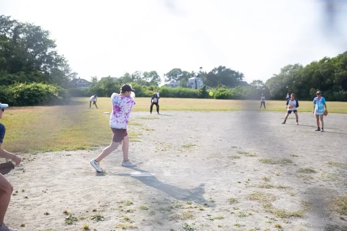
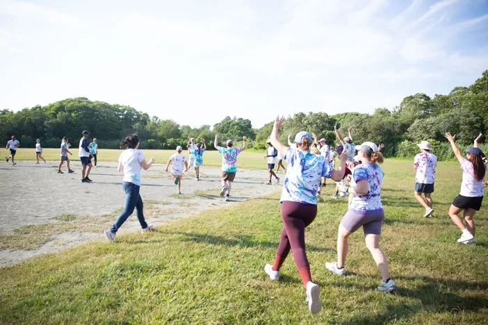
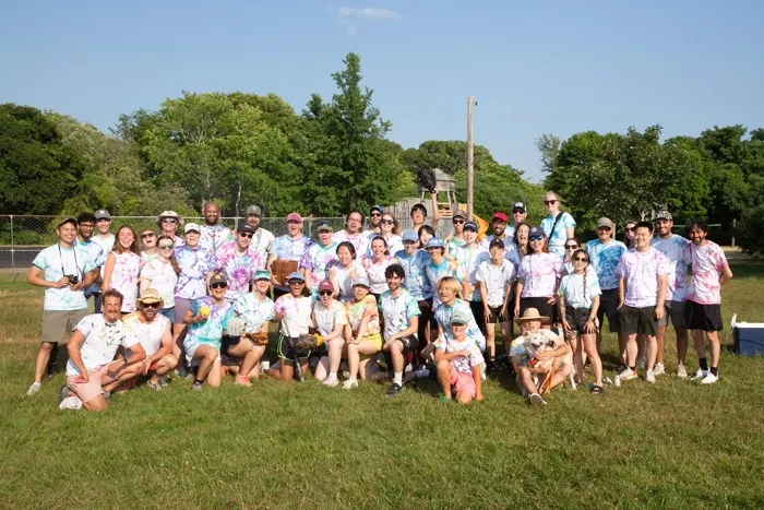
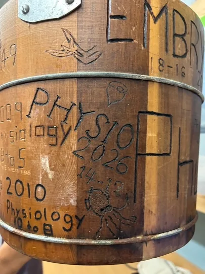
Final remarks
In the middle of Rotation 2, I was still struggling with mounting embryos and looked quite upset in the sample-prep room. Someone from my rotation group asked me, “Why are you so serious, Tingran?” But I was not the only one taking it very seriously. Although the purpose of Physio is to have fun, many people felt a great deal of pressure and self-doubt. (One day, the dog-food-serving dining hall played Queen’s “Under Pressure,” and we were like, “This is the song of the course.”) People were really hard on themselves. That pressure certainly crushed me during Rotation 1. My girlfriend came to see me, and we went back to Boston on Sunday. She pulled me into seeing a random ballet movie called Black Swan. The plot was about a ballet dancer placing impossibly high demands on herself while practicing for Swan Lake, which eventually led to her metamorphosis into a black swan. After watching this, I told my girlfriend that I should maybe also lower the standards I set for myself. After all, I am a theorist. But once I returned to MBL, the environment - the aura created by the people - immediately dispelled any thought of slacking off. The motivation would not fade. For me especially, another reason for driving myself like a machine was that the wet-lab experiments I did at Physio might be the last wet-lab experiments I would do for the rest of my life. I may do experiments during my postdoc, but who knows whether I will even do a postdoc or not? Seize the day! I knew that some people, including me, began to feel quite unwell (e.g., heart discomfort) toward the end because of the continuous sleep deprivation.
The purpose of elaborating on the stress is not to elicit sympathy. In fact, we were truly privileged to have the chance to be so immersed in pure science for 6 weeks. The end of the course meant returning to reality for many of us - failing projects, publishing papers, looking for postdocs, and all kinds of responsibilities. For those 6 weeks, however, we had the golden chance to begin independent work directly on projects whose scientific mysteries were still largely unresolved. Some projects had the potential to have huge impacts, and Physio served as the “testing ground” for them. For example, in 2008, Jonathan Weissman brought the Ribo-seq prototype to here, which went on to revolutionize how we study translation starting in 2009. However, the preparatory work, the heavy-lifting - e.g., making cell lines, protocol optimization, protein purification, and quality control - had already been done long before the course began. Physio felt like pure, unconstrained scientific play only because enormous amounts of labor, money, preparation, and collective pressure had temporarily insulated us from ordinary academic life. In Lucas Pelkmans’s rotation group, the TA spent 3 months purifying DRK3, and they brought all the aliquots they had made for the students in their group to microinject into oocytes. Most TAs also stayed up super late with us every day to support us or, in my case, to babysit me. Some of us also had a chance to use some genuinely remarkable instruments, thanks to the culture MBL had created, which encouraged companies to test their state-of-the-art instruments there. Many of us got to use arguably the best commercial super-resolution microscope for a week (from CSR). One team in Hernan’s rotation group used a $500,000 embryo-sorting machine for its high-throughput screening experiments. Given the increasingly difficult funding situation, this was even more precious. (The national research-funding cuts had also shortened this program by 1 week. Next year, it would be shortened by yet another week. It is worrying whether Physio and the entire MBL can remain financially sustainable in the near future. Some of their ACs have not been repaired for years.)
These 6 weeks felt long. Much longer than 6 weeks. MBL felt like Der Zauberberg (The Magic Mountain) of science. In Magic Moutain, the protagonist, Hans Castorp, went into an Alpine sanatorium to look after his cousin who had TB. He planned to stay there for 3 weeks, but he stayed there for 7 years. During these 7 years, he was totally immersed in art and philosophy. Time was very non-linear in this timeless bubble. He finally stepped out of the sanatorium only to be met by World War I, which we all knew what happened. This was somewhat reminiscent of how I felt after the course ended. When I just went back to Boston, it felt like being an apparition in the crowd. There was no real sense of “today is July 26th”, because I could not even tell how much time had flown past during Physio. Part of me has been forever left at MBL. I also quickly learned about the news that GPT 5.6 had just cracked modern mathematical sciences. We might have also just witnessed the last human-awarded Fields Medal. Science is under unprecedented threat – or progress, who knows – from agentic AI as well – a sentiment that was echoed throughout the course. Exciting times.
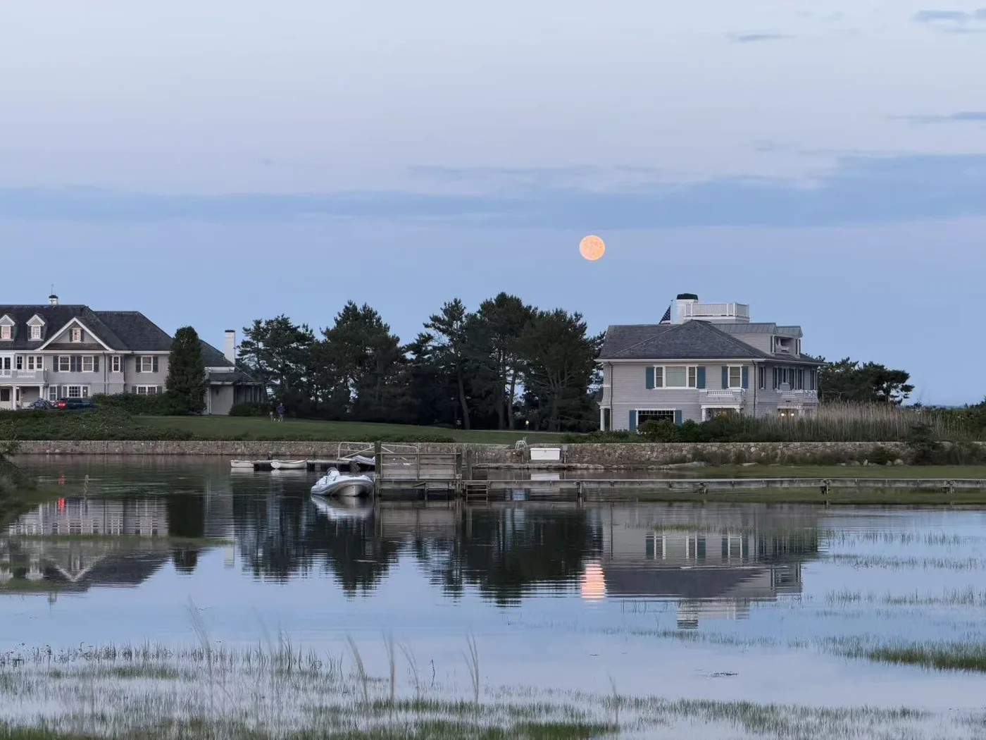
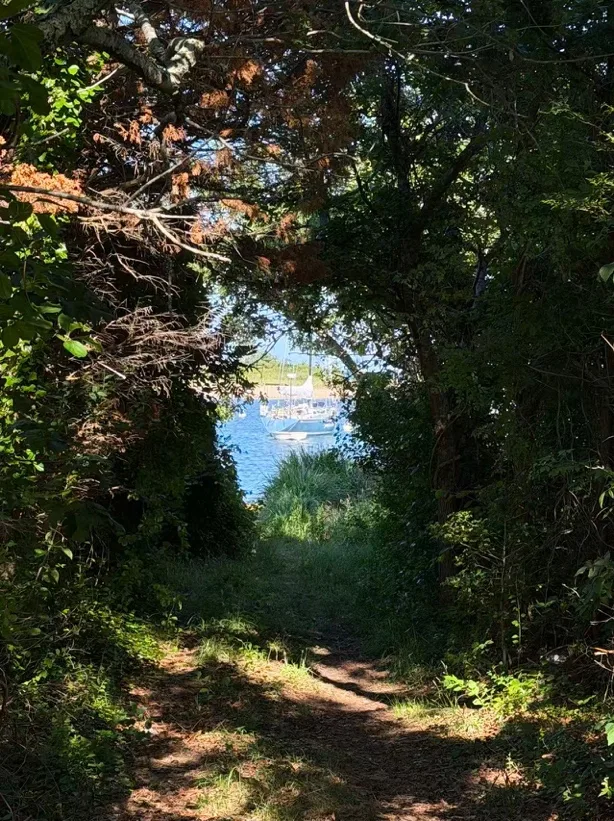
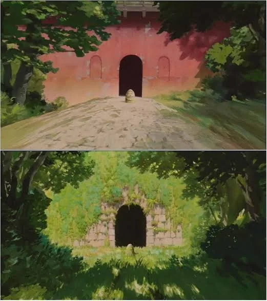
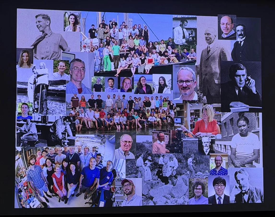
One month later, at the end of summer, my group at MIT returned to MBL for our 25th-anniversary retreat. We invited alumni and friends from all over the world for a small conference and reunion. As the youngest generation there, I loved hearing the histories behind our past science, especially from the people who had made it more than a decade ago: the “stories behind the scenes,” and portraits of the now-veterans when they were still young. I was also surprised by how some past MBL group retreats, brief as they were, had been great sources of inspiration and had sparked future major works.
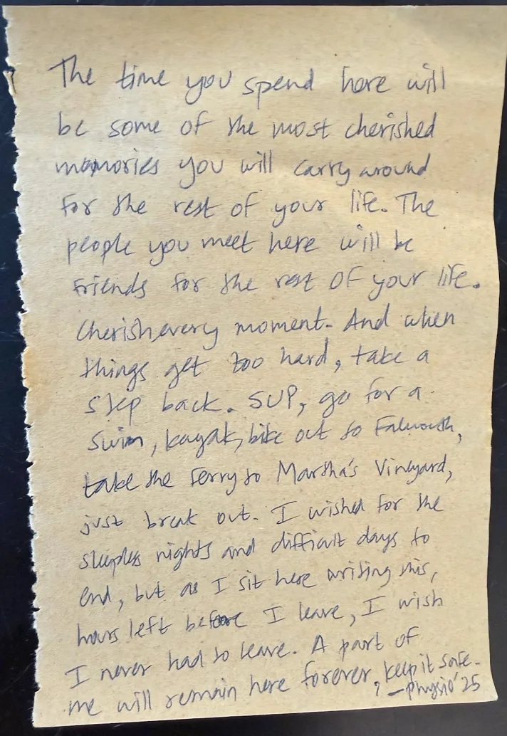
Indeed, this place is truly a place for high creativity, for being intellectually stimulated. When the old crew returned to MBL and began talking about chromosomes and everything else, it felt as though no one had ever truly stepped away from science, whatever they were doing now. There is a real gravity. As one of the retreat speakers remarked, “science is best done with friends.” If I were to return to MBL 20 years later, the influence of Physio would not have faded away, not only because of the science, but because of the friends we met, and the fun we experienced. Time will fly and people will scatter around the globe eventually, but the memories will stay. It will always be our home sweet home.